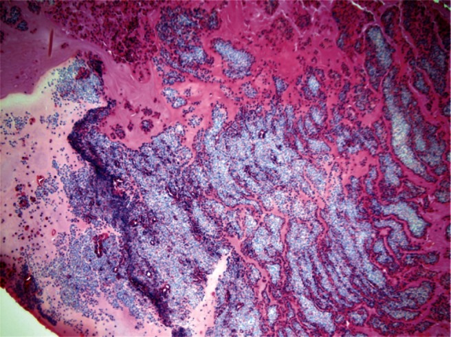
Figure 2.

Hematoxylin and eosin stain (100× magnification) showed inspissated mucin containing collections of chronic inflammatory cells in a laminated arrangement. Focal aggregates of eosinophils are present. A special stain for fungi shows fungal elements morphologically consistent with Aspergillus or Bipolaris species.
