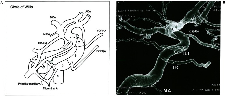
Figure 5.
A) Intra cranial segments (4 to 7) successively extending between the mandibular (not seen) and the trigeminal-primitive maxillary arteries, the dorsal ophthalmic artery, the ventral ophthalmic artery and the bifurcation. B) internal carotid angiography with the 5th cranial segments arterial boundaries visible: Mandibular artery MA, trigeminal remnant TR, Infero lateral trunk ILT, ophthalmic artery OPH.

