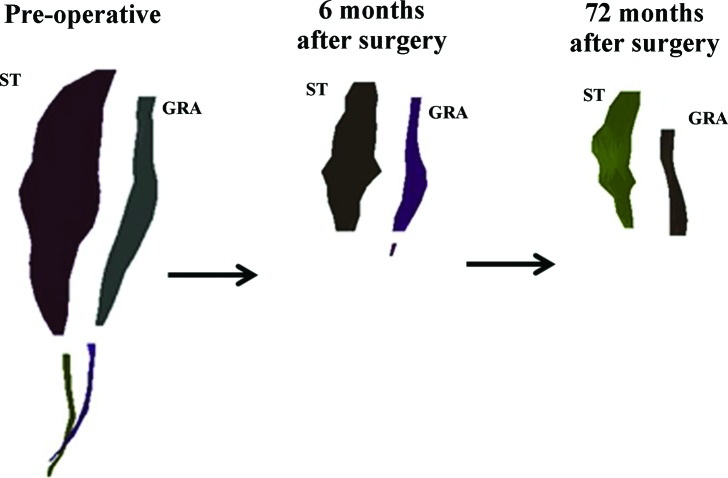
Figure 2.
A three dimensional surface mesh of the semitendinosus (ST) and gracilis (GRA) muscles from the involved limb that was produced by digital reconstruction of the magnetic resonance images of case 1, taken over the three time points demonstrates the poor regeneration of these muscles. Muscle and tendon contours are separated to clearly distinguish the relative morphology differences over time.

