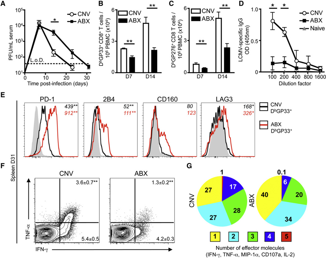
Figure 1. Systemic LCMV T1b Infection Results in Delayed Viral Clearance and an Impaired LCMV-Specific CD8+ T Cell Response in ABX Mice.
(A) Viral titer in the serum of CNV or ABX C57BL/6 mice after LCMV T1b infection (L.o.D., limit of detection).
(B and C) LCMV-specific (B) DbGP33 and (C) DbGP276 tetramer+ CD8+ T cells per 106 peripheral blood mononuclear cells (PBMC) at d7 and d14 p.i.
(D) Serial dilution of LCMV-specific IgG antibody titers in serum of CNV or ABX mice at d23 p.i. Naive serum from CNV mice used for baseline.
(E) Expression of inhibitory receptors PD-1, 2B4, CD160, LAG-3 on DbGP33 tetramer+ CD8+ T cells isolated from the spleen of CNV (black line) or ABX (red line) mice. Shaded histograms represent CD44lo CD8+ T cells. Numbers in italics represent mean fluorescence intensity (MFI).
(F and G) Splenocytes from d31 infected mice were incubated with GP33 peptide for 5 hr in the presence of BFA and assessed for production of IFN-γ, TNF-α, MIP-1α, CD107a, and IL-2. FACS plots gated on live, CD8α+ cells.
(G) Proportion of GP33 peptide responsive CD8+ T cells producing multiple effector molecules. Data representative of three independent experiments with n = 5 mice per group. Data shown are the mean ± SEM. Serum viral titer statistics determined by two-part t test for each time point. *p < 0.05, **p < 0.01. See also Figure S1.

