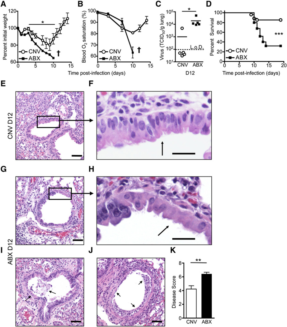
Figure 2. Alterations in Commensal Bacterial Communities Exacerbate Lung Pathology and Mortality to Influenza Virus.
CNV or ABX C57BL/6 mice were infected i.n. with influenza virus PR8-GP33.
(A and B) Time course of weight loss (A) and blood oxygen (B) saturation after infection (representative exp. n = 5: † signifies mice below 70% initial weight were sacrificed).
(C) Influenza virus genome copies in the lung at d12 p.i. assessed by RT-PCR and displayed as TCID50/gram of lung tissue.
(D) Survival curve after PR8-GP33 infection; CNV n = 27, ABX n = 25.
(E–J) H&E-stained lung section of CNV (E and F) or ABX (G–J) mice at d12 p.i. Black box and arrows highlight (E and F) epithelial hyperplasia, (G and H) epithelial cell necrosis, (I) cellular debris and exudate in lumen, and (J) loss of bronchiole epithelium (scale bar represents 50 µm in E, G, I, and J; 20 µm in F and H).
(K) Disease score of bronchiole epithelial degeneration at d12 p.i. Data representative of five independent experiments with n = 5–6 mice per group. Survival statistics determined by log rank test. Viral titer statistics determined by two-part t test. *p < 0.05, **p < 0.01, and ***p < 0.001. Data shown are mean ± SEM. See also Figure S2.

