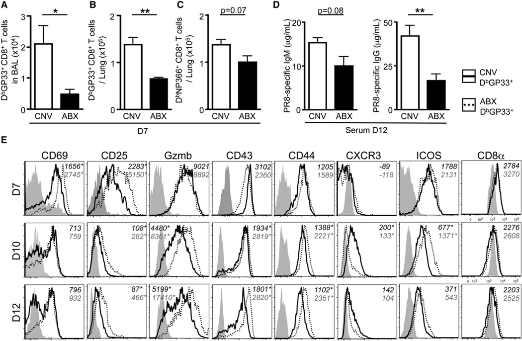
Figure 3. ABX Mice Have a Diminished Influenza Virus-Specific Adaptive Immune Response.
(A and B) Total number of influenza virus-specific DbGP33 tetramer+ CD8+ T cells isolated from the (A) BAL or (B) lung parenchyma at d7 p.i.
(C) DbNP366 tetramer+ CD8+ T cells isolated from the lung parenchyma at d7 p.i.
(D) PR8-specific IgM and IgG titers in the serum at d12 p.i.
(E) Phenotypic profile of DbGP33 tetramer+ CD8+ T cells isolated from the lung of CNV (solid line) or ABX (dotted line) mice at d7, d10, and d12 p.i. Gray shaded histograms are CD44lo CD8+ T cells isolated from the lung. Numbers in italics represent MFI. Data representative of three independent experiments with n = 4–5 mice per group. *p < 0.05 and **p < 0.01. Data shown are mean ± SEM. See also Figure S3.

