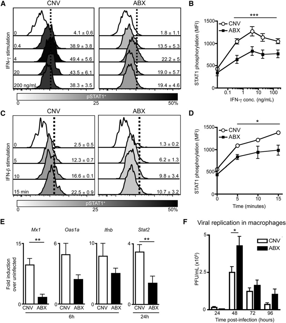
Figure 6. Macrophages from Naive ABX Mice Have a Diminished Ability to Respond to IFN Stimulation and Viral Infection In Vitro.
(A–D) Peritoneal Macrophages isolated from CNV or ABX mice were stimulated with IFN-γ or IFN-β in vitro. Histograms of STAT1 phosphorylation in macrophages after (A) IFN-γ stimulation (0.4, 4, 20, 200ng/mL) or (C) IFN-β stimulation (103 units/mL for 5, 10, and 15 min). MFI of pSTAT1 in macrophages after (B) IFN-γ or (D) IFN-β stimulation.
(E and F) Peritoneal macrophages sorted from naive CNV or ABX mice were infected in vitro with (E) influenza virus (X31-GP33, MOI of5)or(F) LCMV (cl-13 strain, MOI of 0.2).
(E) Induction of antiviral defense genes in macrophages at 6 and 24 hr p.i. as assessed by RT-PCR.
(F) LCMV viral titers in supernatant at 24–96 hr p.i. Data representative of two or more independent experiments with n = 3–5 mice per group. *p < 0.05, **p < 0.01, and ***p < 0.001. Data shown are mean ± SEM. See also Figure S7.

