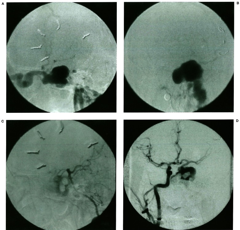
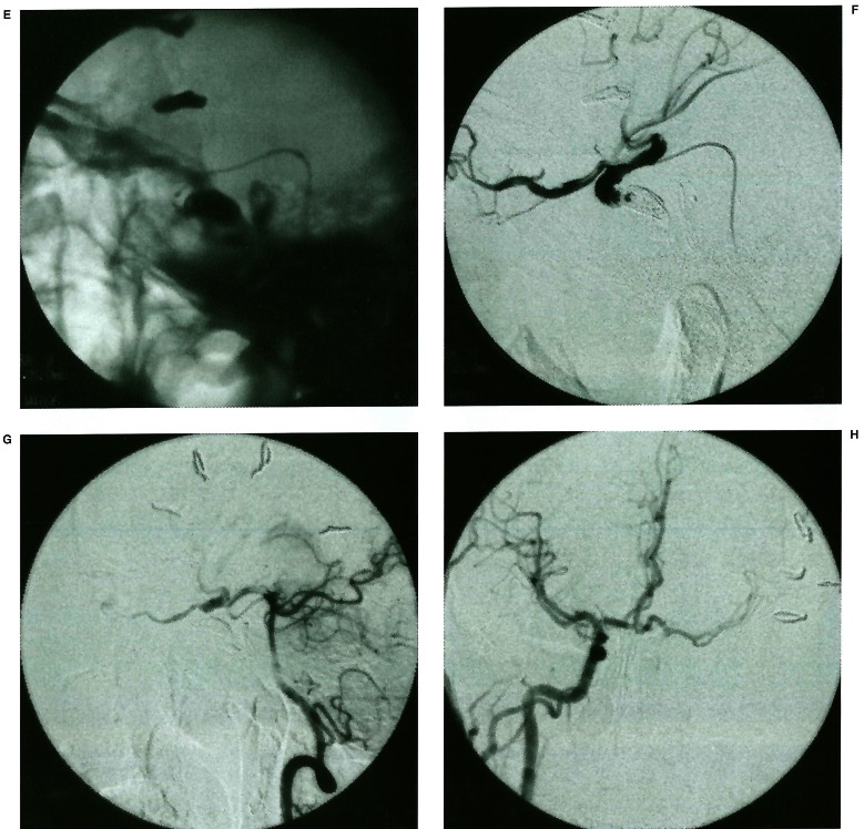
Figure 3.
A 26-year-old male suffered of TCCF with severe epistaxis. Angiogram could show the sign of pseudoaneurysm protruding into sphenoid sinus (A, B). Trying to occlude internal carotid artery could not embolise the fistula completely and epistaxis could not be stopped. Fistula was supported by anterior and posterior communicating artery (C, D). The microcatheter was introduced into fistula through posterior communicating artery. The fistula was totally embolised by MDS (E, F). Contralateral carotid artery and vertebral artery angiogram showed the good result of embolisation (G, H).


