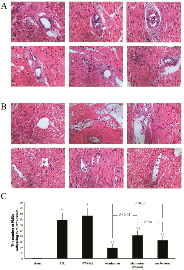
Figure 2.
Assessment of PMN adherence to microvessels by H&E staining. A) Micrographs showing PMNs adhering to arterioles: (a) sham-operated group; (b) I/R group; (c) GW9662 group; (d) telmisartan group; (e) telmisartan–GW9662 group; (f) candesartan group; (H&E stain; 400×). B) Micrographs show PMNs adhering to venules: (a) sham-operated group; (b) I/R group; (c) GW9662 group; (d) telmisartan group; (e) telmisartan–GW9662 group; (f) candesartan group; (H&E stain; 400×). C) Quantitative analyses of the number of PMNs adhering to microvessels. *P < 0.05 compared with sham-operated; #P < 0.05 compared with I/R.

