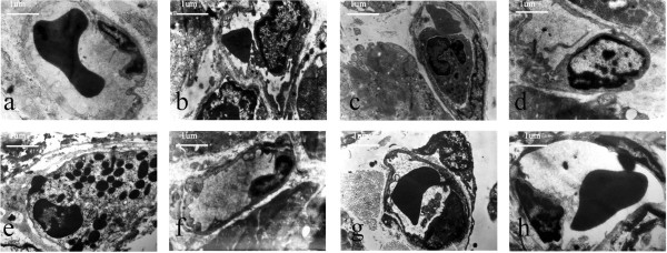
Figure 4.

Photomicrographs of the ultrastructural changes in rabbit capillaries. (a) sham-operated group, capillary basement membrane was moderate in thickness, and the capillary contained an erythrocyte (7700×); (b) I/R group, endothelial cell swelling led to stenosis of the capillary lumen (7700×); (c) I/R group, one leukocyte plugged a single capillary (7700×); (d) GW9662 group, endothelial cell swelling led to stenosis of the capillary lumen (7700×); (e) GW9662 group, one leukocyte plugged a single capillary (7700×); (f) telmisartan group, endothelial cell swelling was relieved (7700×); (g) telmisartan–GW9662 group, endothelial cell swelling was relieved (7700×); (h) candesartan group, endothelial cell swelling was relieved (7700×).
