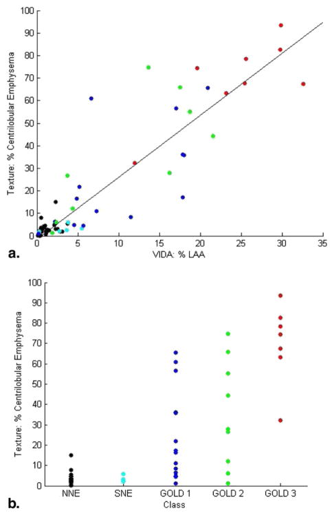
Figure 5.
Percentage emphysema obtained using the texture-based method is correlated with (a) percentage low-attenuation area (LAA) computed by the density mask technique and (b) chronic obstructive pulmonary disease (COPD) Global Initiative for Chronic Obstructive Lung Disease (GOLD) stage. Black data points represent scans of normal subjects with no emphysema (NNE); cyan represents scans of smokers with no emphysema (SNE); and blue, green, and red represent GOLD stages 1, 2, and 3, respectively. The differences in percentage emphysema computed using the texture-based method were statistically significant (P < .001) by analysis of variance. SNE scans were not statistically significantly different from NNE scans, but GOLD stages 1, 2, and 3 were statistically significantly different from both NNE and SNE classes (P < .05).

