
Fig. 1.
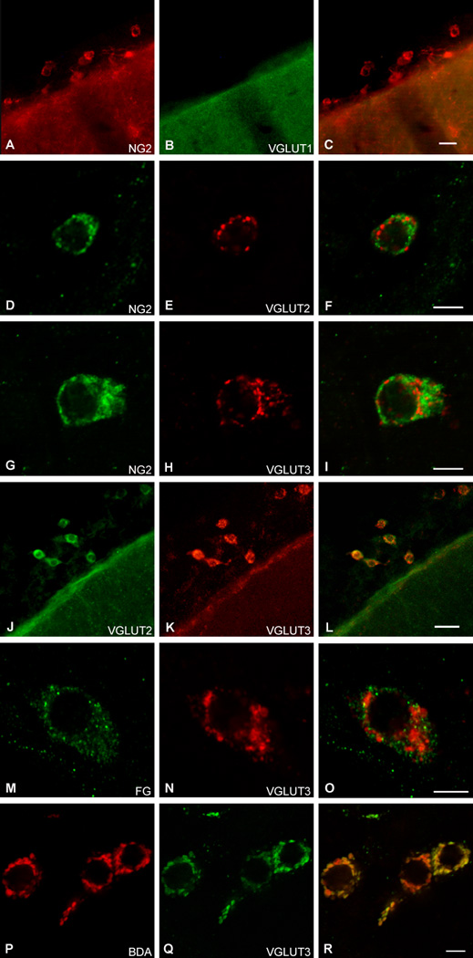
Leptomeningeal CNS pericytes co-express VGLUT2 and VGLUT3 in vesicles. VGLUT1 is not expressed in CNS pericytes (revealed by NG2 proteoglycan (NG2)-ir) (A–C). However, both VGLUT2 (D–F) and VGLUT3 (H–J) are expressed in CNS pericytes, where they appear to be co-localized (J–K). VGLUTs were not seen in lysosomes as revealed using FluoroGold (FG) as a functional marker of these organelles (M–O). Instead,VGLUT3 was localized to secretory vesicles as revealed by the co-localization of the transporter with biotinylated dextran amine (BDA), which is taken up into vesicles from the extracellular environment during exocytosis (P–R). Antibodies: (A–C) mouse anti-VGLUT1; (D–F) guinea pig anti-VGLUT2; (J–L) rat anti-VGLUT2. Scale bars: (C, L) 20 µm; (F, I, O, R) 5 µm. *Blood vessel.