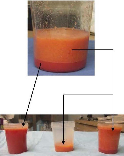
Figure 1.
Images of standard lipoaspirate (top container) after Coleman technique separated into aqueous phase (far left container) and yellow appearing low SVF fat (center container) and orange appearing high SVF (right container). (Oil phase not pictured.) Because the fat is already pre-washed, the heme pigmentation is not a result of contaminated red blood cells (RBCs), but rather RBCs that are still within the perivascular space thereby creating an indirect marker for a fat fraction rich in perivascular cells (i.e. orange appearing fat).

