
Figure 3.
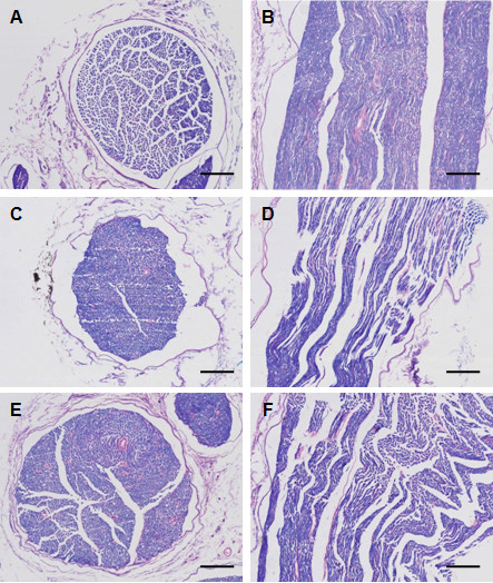
Histological examination of luxol fast blue stained sciatic nerve. (A, B): Normal control; (C, D): Diabetic peripheral neuropathy (DPN); and (E, F): High-dose rhTNFR:Fc (4 mg/kg) group (DPN + T2). Left panel shows cross-sections with 100X amplification (A, C, E, scale bar = 150 μm), and right panel shows longitudinal sections with 300X amplification (B, D, F, scale bar = 50 μm).