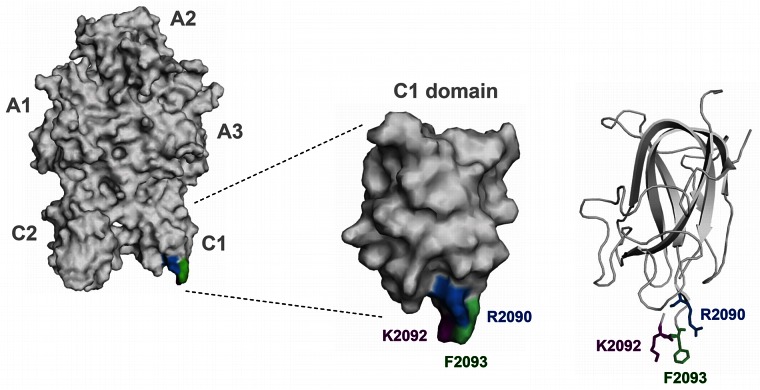
Figure 1.
Three-dimensional structure of FVIII molecule highlighting C1 domain residues Arg2090 (blue), Lys2092 (purple), and Phe2093 (green). Left panel provides an overview of the entire FVIII molecule. The position of the A1, A2, A3, C1, and C2 domains is indicated next to the model. The middle panel provides an overview of the surface coverage of the C1 domain. The right panel shows the secondary structure of the protein backbone of the C1 domain. Residues Arg2090, Lys2092, and Phe2093 are displayed in a ball and stick format. Models were based on FVIII crystal structure (PDB code 3cdz) and prepared with PyMol V0.99 imaging software (Schrodinger).

