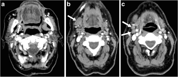
Figure 1.
Axial contrast enhanced CT images of the neck in an 81 year old male (Patient Z in Table 3); a fine needle aspiration biopsy of a clinically enlarged right submandibular lymph node had shown squamous cell carcinoma. (a) A plaque-like (2.5 cm in diameter × 0.5 cm in maximum thickness) enhancing mucosal lesion involving the right retromolar trigone (arrow) was not mentioned in the original radiology report but was identified as the primary tumor in the second opinion report. (b) An enlarged (2.3 cm × 1.5 cm) right level IB lymph node (arrow) was interpreted as metastatic in both the original and second opinion reports. (c) Two sub-centimeter but rounded and asymmetrically prominent right level IIA lymph nodes (arrows) were not mentioned in the original report but were interpreted as metastatic in the second opinion report. Staging was TxN1 based on the original report and T2N2b (Oral Cavity) based on the second opinion report, and the recommended management was ‘don’t know’ based on the original report (because the primary tumor location and T-category were unclear) and ‘surgery’ based on the second opinion report. Surgery was subsequently performed and the final pathologic staging was T2N2b (Oral Cavity).

