Figure 3.
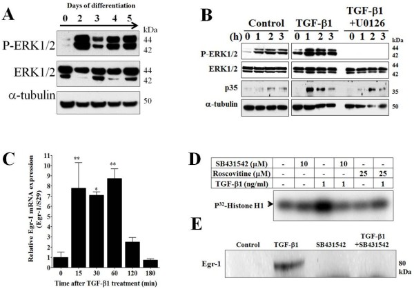
Differentiation or TGF-β1 treatment induces activation of the ERK1/2 pathway and increases Cdk5 activity in MDPC-23 cells. A, Representative Western blot analysis showing activation of ERK1/2 signaling pathways measured by an increase in phospho-ERK1/2 on successive days during differentiation of MDPC-23 cells. B, Representative Western blot analysis of phospho-ERK1/2, ERK1/2, p35, and α-tubulin was performed in MDPC-23 cells treated with: vehicle (control), TGF-β1 (1 ng/ml), and TGF-β1 (1 ng/ml) plus U0126 (20 μM) over the course of 0, 1, 2, and 3 h. C, qPCR analysis of Egr-1 mRNA levels normalized against S29. Total RNA was obtained from MDPC-23 cells treated with TGF-β1 (1 ng/ml) for 0, 15, 30, 60, 120, and 180 min. D, The effect of roscovitine on Cdk5 kinase activity. MDPC-23 cells were treated with vehicle (control), SB431542 (10 μM), TGF-β1 (1 ng/ml), TGF-β1 (1 ng/ml) plus SB431542 (10 μM), roscovitine (25 μM), and TGF-β1 (1 ng/ml) plus roscovitine (25 μM) over 24 h. E, Western blot analysis against Egr-1 from MDPC-23 cells treated with: vehicle (control), TGF-β1 (1 ng/ml), SB431542 (10 μM), and TGF-β1 (1 ng/ml) plus SB431542 (10 μM) over 24 h. All data are presented as the mean and SEM (n = 4). *p < 0.05, **p < 0.01, (Dunnett’s test after ANOVA).

