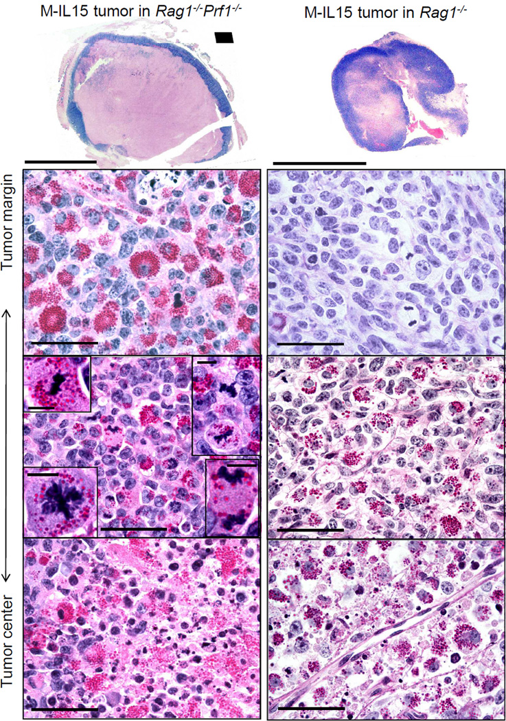
Figure 5. Eradication depends on eliminating cancer at the tumor rim.
Macroscopic and microscopic photographs of tumors grown in anti-IL15 treated mice. Left, a Rag1−/−Prf1−/− mouse 31 days after M-IL15 injection and 14 days after the final anti-IL15 injection. Right, a Rag1−/− mouse at 16 days after M-IL15 injection and 2 days after final anti-IL15 injection. Tumor sections were stained with PAS & diastase. Scale bars for macroscopic photographs are 1cm. Microscopic photographs were taken in various regions of the tumors, as depicted on the left. Scale bars for microscopic photographs are 100µm. Insets show mitotic granular NK cells (scale bars are 10µm).

