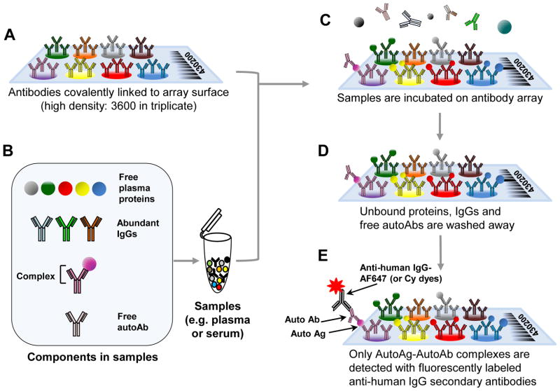
Figure 1. A schematic overview of the autoantibody-autoantigen complex profiling array method.
(A) Antibodies are spotted onto slides and covalently linked via N-hydroxysuccinimide (NHS)-ester reactive groups to the slide. (B) The sample containing the autoantibody complexes, free plasma proteins and IgGs is applied to the array. (C) Proteins in the serum or plasma samples are affinity captured when the appropriate commercial antibody is present on the antibody microarray surface. (D) Only plasma proteins that bind tightly to the capture antibodies on the array will remain after washing. (E) The autoantibody-antigen complex is detected by fluorescently tagged, anti-human secondary antibody.

