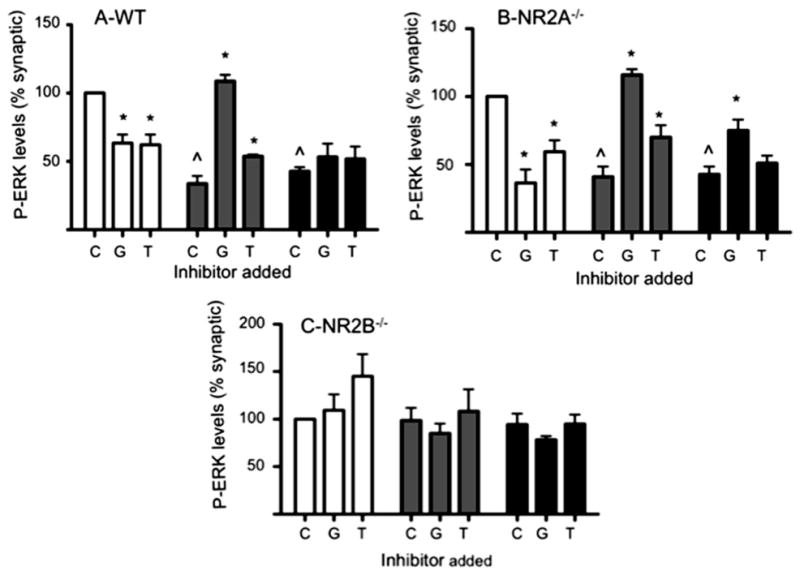
Fig. 3.

NR2B is required for extrasynaptic activation. Quantification of the effect of 40 μM con-G (G) and 40 μM con-T (T) on P-ERK1/2 levels with DIV 13–15 mouse cortical neurons, along with an untreated control (C). A. WT mouse cortical neurons. B. Mouse cortical NR2A−/− neurons. C. Mouse cortical NR2B−/− neurons. The neurons were treated to activate synaptic (white bars), extrasynaptic (gray bars), or total (black bars) NMDARs. P-ERK levels are relative to synaptic activation, taken as 100%. Quantitation of P-ERK staining shown as mean ± SEM from three independent experiments. *p < 0.05, compared with a control (C) within each group. ^p < 0.05, compared with the synaptic control (C).
