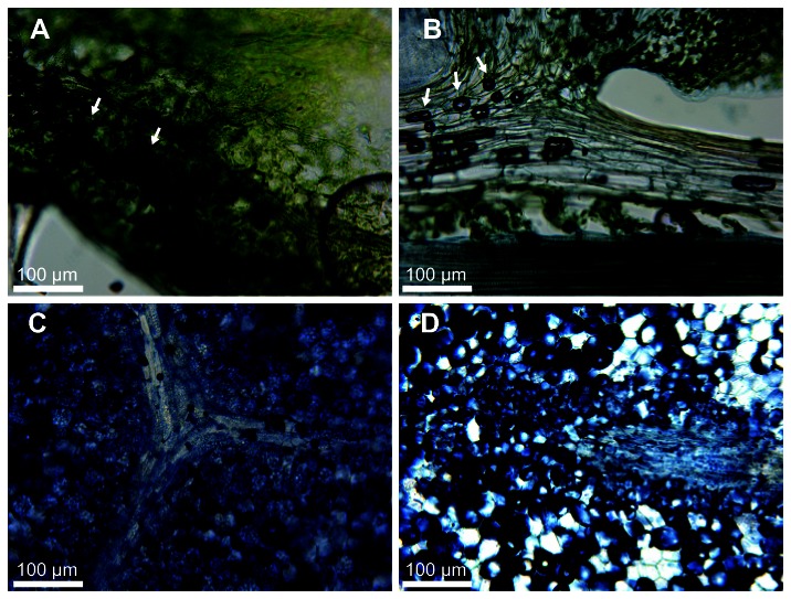
Figure 5. Lipid storage in bulbils.
Vibratome-cut sections were stained with Sudan Black B to identify cells with neutral lipids. (A) Section through an Arabidopsis leaf with a low storage lipid content. (B) Stem and leaf section from S . moellendorffii . (C) Section through a pumpkin oilseed with high storage lipid content. (D) S . moellendorffii bulbil section. Stained lipids appears as dark blue or black. Air bubbles in A and B (arrows) appear dark and do not indicate staining. The images are representative of material from at least three different plants.

