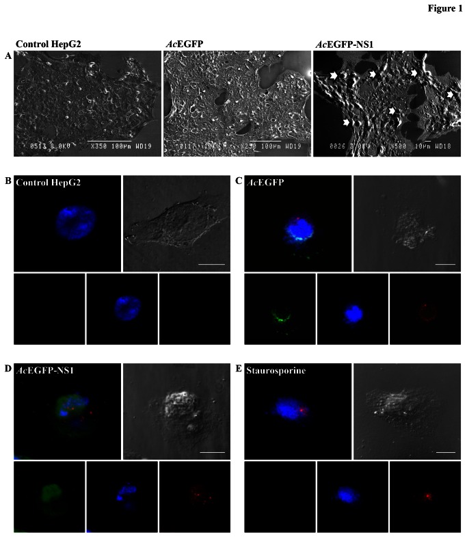
Figure 1. Human Parvovirus B19 NS1 protein induces apoptotic blebs and bodies in non-permissive cell line.
(A) Scanning electron microscopic images of non-infected cells, cells transduced with AcEGFP and AcEGFP-NS1, respectively. ApoBods with potential self-antigens are detected at 48 h post-transduction as indicated with arrows. Bars 100 µm or 10 µm. (B–E) Apoptotic blebs created from NS1 expression show positively for Annexin V-PE. Laser scanning confocal microscopy images of (B) non-transduced HepG2 cells, (C) cells transduced with AcEGFP, (D) cells transduced with AcEGFP-NS1, and (E) staurosporine treated cells. Cells were visualized (lower panels) directly for EGFP (green), stained for DNA with DAPI (blue), and labeled for phosphatidylserine (PS) with Annexin V-PE (red). Upper panels represent the merged image of the bottom panels and the DIC micrograph to see the morphology of the surface of the cell. Bars 20 µm.

