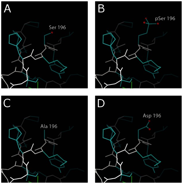
Figure 2. Structural context of phosphorylation and substitutions at EVI1 S196.

. Main chain is shown in white and side chains in blue. Oxygen atoms on residue 196 are indicated by red spheres. (A) The zinc finger domain with unmodified serine, (B) phosphorylated serine, (C) aspartate substitution and (D) alanine substitution. All residues can adopt a similar conformation and extend into the solvent.
