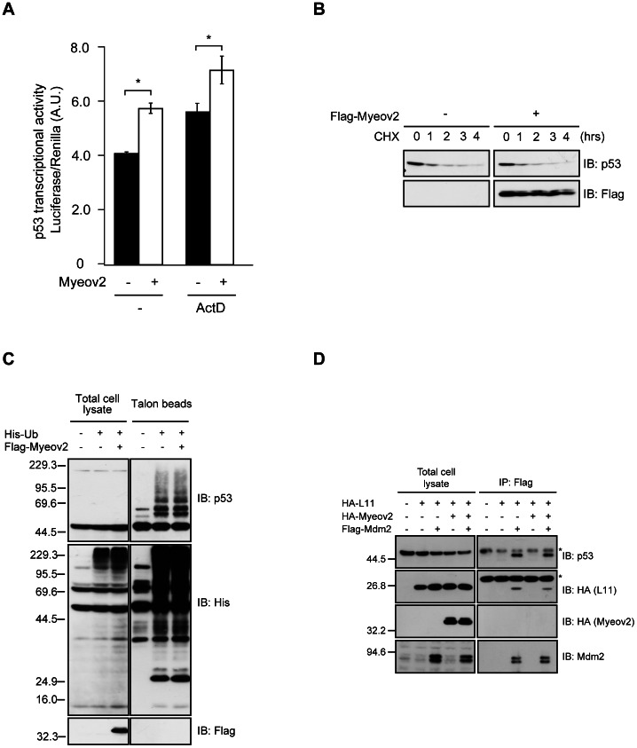
Figure 4. Expression of Myeov2 augments p53 activity independently of ubiquitination.
(A) HCT116 cells were transfected with either Myeov2 or empty vector, together with p53-promoter-luc plasmids, and were then stimulated with or without 5 nM ActD for 6 hours. p53 promoter activity was analyzed by luciferase assay (n = 3, mean±SEM, * p<0.05 by one-way ANOVA). (B) HEK293 cells were transfected with Flag-Myeov2 and were then treated with 10 µM cycloheximide for indicated times. Cell lysates were subjected to immunoblot analyses using anti-p53 and anti-Flag antibodies. (C) HEK293T cells were transfected with indicated plasmids. Ubiquitinated proteins were precipitated from denatured cell lysates using Talon metal affinity resin and immunoblotted with anti-His, anti-Flag, and anti-p53 antibodies. (D) Co-immunoprecipitation of HA-L11, HA-Myeov2 and endogenous p53 with Flag-Mdm2 in HEK293T cells. Cell lysates were immunoprecipitated with anti-Flag antibody and analyzed using Flag, HA, Mdm2 and p53 antibodies. Asterisks indicate non-specific bands.

