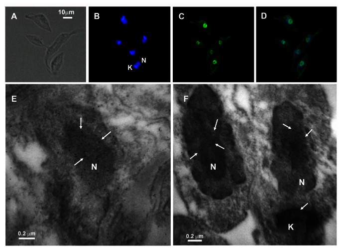
Figure 3. Immunolocalization of PARG on Trypanosoma cruzi, CL Brener epimastigotes.
The parasites were fixed for 25 min with 3.8% (W/V) formaldehyde in PBS at 4°C, permeabilized with fresh PBS - 0,1% Triton X-100 and blocked for 1 h at room temperature with 5% (W/V) BSA in PBS. (A) Differential interference contrast (DIC). (B) Cells were counterstained with DAPI to identify nuclear DNA and kinetoplastid DNA. (C) PARG was detected with 1:500 mouse polyclonal TcPARG antibody followed by 1:600 Alexa Fluor 488 goat anti-mouse IgG antibody. (D) Merge of PARG and DNA signals show the nuclear localization of this enzyme. Bar: 10 µm. (E–F) For electron microscopy, epimastigotes were fixed in PBS 2.5% glutaraldehyde, 4% formaldehyde, embedded in epoxy resin and PARG detected with 1:50 mouse polyclonal TcPARG antibody followed by 1:100 anti-mouse antibody conjugated with 10-nm gold particle. N: nucleus; K: kinetoplast. Bar: 0.2 µm.

