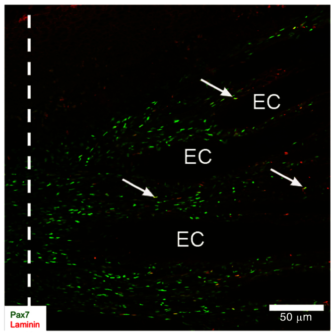
Fig. 5.

Activation of Pax7-positive cells near intact electrocytes (EC). Portions of longitudinal cryosection (20 mm thick) from 14-day blastema co-labeled with anti-Pax7 (green) and anti-BrdU (red) antibodies. White dashed line shows the site of tail amputation. Arrows point to cells that were co-labeled with Pax7 and BrdU. Reproduced from Weber et al. (Weber et al., 2012).
