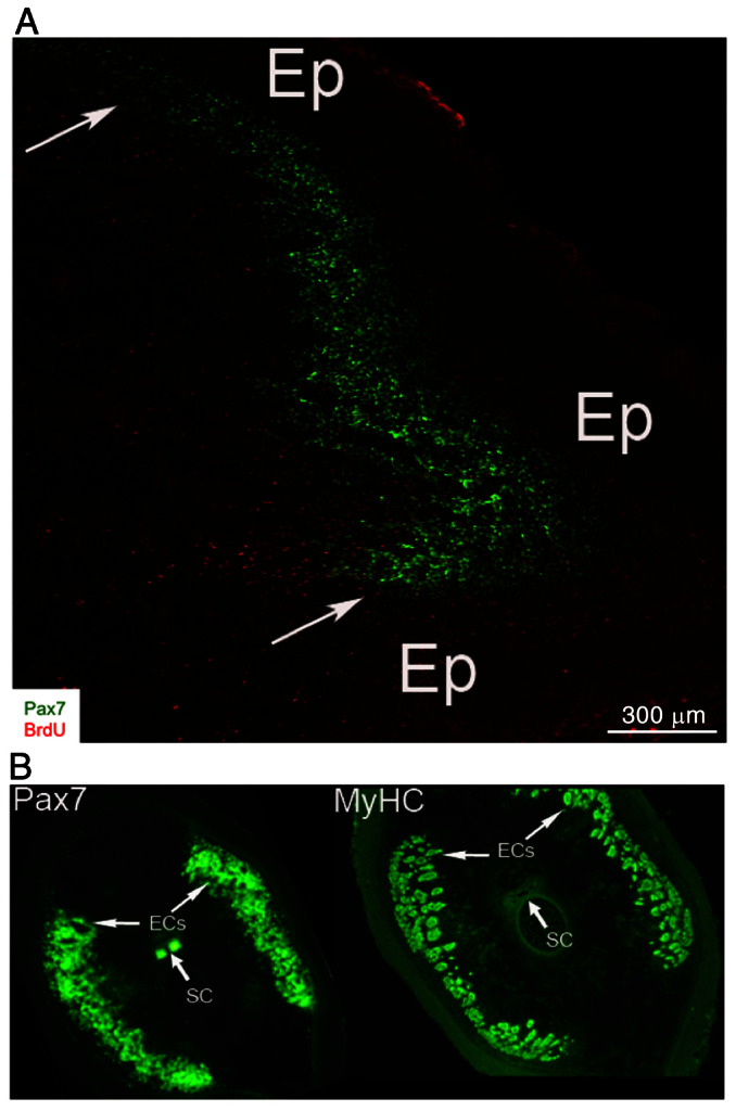
Fig. 6.

Distribution of Pax7-positive cells in regions that give rise to muscle fibers and electrocytes. (A) Spatial distribution of Pax7-positive cells in regeneration blastema. Confocal images of longitudinal cryosection (20 mm thick) from 14-day blastema immunolabeled with anti-Pax7 and BrdU antibodies. Arrowheads point to Pax7-positive cells adjacent to the epithelium (Ep). Reproduced from Weber et al. (Weber et al., 2012). (B) Pax7-positive cells are localized in blastema regions that give rise to muscle, electric organ and dorsal spinal cord. Serial cross-sections taken from distal half of 14-day blastemas immunolabeled with antibodies against Pax7 (green) and myosin heavy chain (MyHC). MyHC is present in all muscle fibers, which are small cells located between epithelium and developing electrocytes (ECs), which are larger cells more medially located (arrows). Co-labeling by anti-Pax7 and anti-MyHC antibodies was detected in peripheral regions of the blastema underneath the epithelium. Pax7 labeling was also detected in dorsal spinal cord (SC). Reproduced from Weber et al. (Weber et al., 2012).
