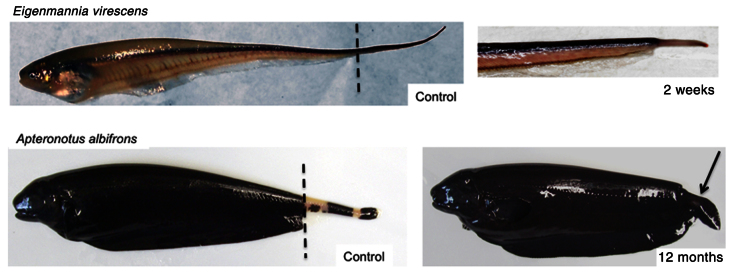
Fig. 8.

Tail regeneration in adult gymnotiforms Eigenmannia virescens and Apteronotus albifrons following amputation. Left panels: adult control Eigenmannia virescens (glass knifefish) and Apteronotus albifrons (black ghost knifefish). In both gymnotiforms, regeneration proceeds through the formation of a blastema at the base of the amputation plane (dashed line) and cell differentiation occurs from proximal to distal regions of the blastema. Right panels: regenerated tails in Eigenmannia virescens and Apteronotus albifrons 2 weeks and 12 months after tail cut, respectively. The 2-week regeneration blastema in E. virescens (top right panel) parallels that observed in S. macrurus and its subsequent regeneration results in the restoration of all tissues and structures in proportions comparable to that of an intact adult tail. In A. albifrons, regeneration after amputation proceeds with blastema formation (not shown) and differentiation of anal-like fin structure (arrow) with stunted tail segment. Photo credit: Vincent Gutschick.
