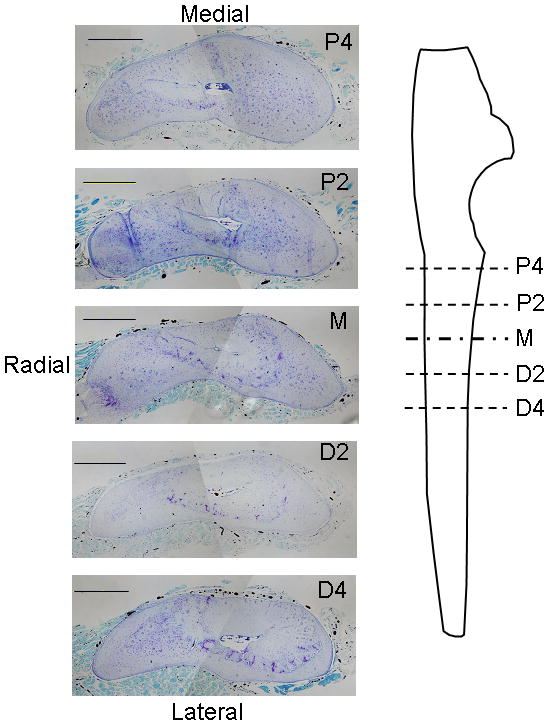
Figure 1.

Photomicrographs of transverse histological sections from a control (left) ulna at five sites along the ulnar length. Perfused vessels were easily visualized as opaque areas within the thin periosteal layer, as well as within bone and muscle. (stained with toluidine blue; scale bar = 500μm) P4: 4 mm proximal to the midpoint; P2: 2 mm proximal to the midpoint; M: midpoint; D2: 2 mm distal to the midpoint; D4: 4 mm distal to the midpoint.
