
Figure 2.

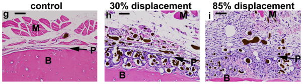
Photomicrographs of transverse histological sections from six loaded (right) and one control (left) ulnae taken 2 mm distal to the midpoint (D2) (orientation same as in Figure 1). (a–f) Low power images of representative samples from the lowest (30%) and highest (85%) displacement groups 3, 7 and 14 days after fatigue loading (10X objective; scale bar = 200 μm). (g–i) Higher power images of ulnae on day 3 (40X objective; scale bar = 50 μm). Loading caused a dramatic expansion of the periosteal and sub-periosteal tissue layers by day 3 for all displacement groups, with greater expansion observed in the higher displacement groups; note that the muscle [M] has been displaced away from the bone [B] in the loaded ulnae compared to control. The number and size of periosteal vessels were greatly increased in loaded ulnae. On day 3, a clear demarcation is seen at higher power (h,i) between the sub-periosteal layer (tissue between the original bone and the periosteal margin [P]) and the periosteal layer (which we define as the tissue between the periosteal margin and the muscle). The sub-periosteal layer is filled with numerous plump cuboidal cells surrounding the blood vessels, and small buds of newly formed bone (pink) are seen; the periosteal layer has a looser, fibrovascular appearance. By day 7, the sub-periosteal layer has transformed to woven bone and the periosteal layer is reduced in size. Isolated regions of chondroid bone (d, *) were observed in the 85% displacement group at this timepoint. By day 14, the woven bone layer has consolidated and the periosteal layer is further reduced. On days 7 and 14, the major site of new vessels is in channels in the woven bone, whereas at day 3 the new vessels are not yet enveloped in bone. (stained with hematoxylin and eosin)