
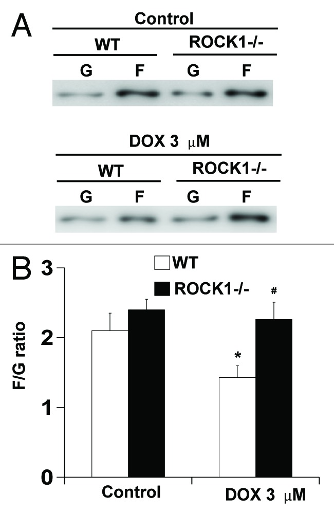
Figure 2. ROCK1 deletion preserves cell F-actin content in response to doxorubicin. WT and ROCK1−/− MEFs with or without 3 μM doxorubicin for 4 h. G- and F-actin were separated by ultracentrifuge followed by western blot of supernatants (G-actin) and pellets (F-actin). Representative western blots (A) were analyzed for F/G-actin ratios (B). *p < 0.05 vs. control of the same genotype; #p < 0.05 vs. WT under the same treatment condition.