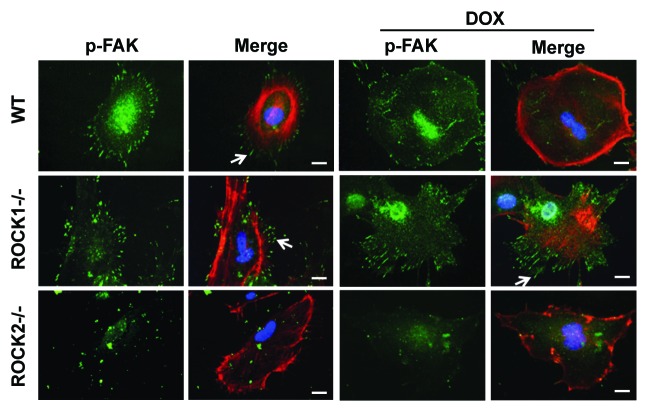
Figure 3. ROCK1 deletion, but not ROCK2 deletion, reduces doxorubicin-induced impairment of focal adhesion formations. Representative images of focal adhesion formation revealed by p-FAK staining (green) of individual WT, ROCK1−/− and ROCK2−/− cells at baseline and after treatment with 3 μM doxorubicin for 16 h. Rhodamine-phalloidin staining for F-actin (red), and DAPI staining for nuclei (blue) are shown in merged images. Typical focal adhesions are indicated with white arrows. Bar, 50 μm.

An official website of the United States government
Here's how you know
Official websites use .gov
A
.gov website belongs to an official
government organization in the United States.
Secure .gov websites use HTTPS
A lock (
) or https:// means you've safely
connected to the .gov website. Share sensitive
information only on official, secure websites.
