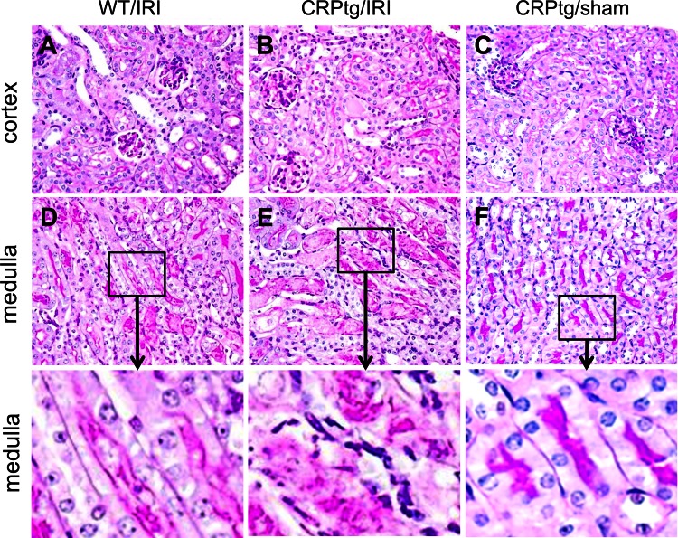
Fig. 6.
Histological changes after renal IRI. Histology [periodic acid-Schiff (PAS) staining] of the renal cortex (A–C) and outer medulla (D–E) of representative kidneys collected from WT and CRPtg mice 24 h after renal ischemia reperfusion injury (IRI) or sham-surgery (sham). Original magnification of A–F is ×40. Bottom: 4-fold magnifications of the areas indicated in D–F. Note the PAS-positive brush border is intact in F and either damaged or lost in D and E. Also note the extensive tubular casts and necrotic cells in E.

