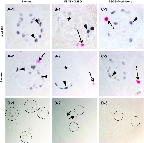
Fig. 5.
Podocyte proliferation measured by p57/Ki-67 double staining. A–C: representative images of p57/Ki-67 staining at 2 and 4 wk at ×630 original magnification. A-1, A-2: normal mice: p57-positive nuclei (blue/gray) are detected in a typical podocyte distribution (arrowheads). No Ki-67 staining is detected in normal glomeruli. A tubular cell stains positive (red, dashed arrow) at 4 wk (A-2). B-1, B-2: mice with FSGS given DMSO: overall fewer p57 staining cells are detected; p57 staining is not detected in areas of sclerosis (*). At 2 wk, Ki-67 staining is detected in a cell lining Bowman's capsule, and at 4 wk positive staining is detected in a tubular cell. C-1, C-2: mice with FSGS given prednisone: at 2 wk, Ki-67 staining is detected in a cell lining Bowman's capsule, and at 4 wk positive staining is detected in a tubular cell. D1–3: negative controls (×200 magnification). D-1: staining for Ki-67 was not detected when the primary antibody was omitted (dashed circles represent glomeruli with p57 staining). D-2: staining for p57 was not detected when the primary antibody was omitted (dashed circles represent glomeruli; arrow represents a positive Ki-67 cell). D-3: staining for p57 or Ki-67 was not detected when both primary antibodies were omitted (dashed circles represent glomeruli). These data show that podocytes do not proliferate in mice with FSGS given DMSO or prednisone. Ki-67 staining is detected in cells lining Bowman's capsule.

