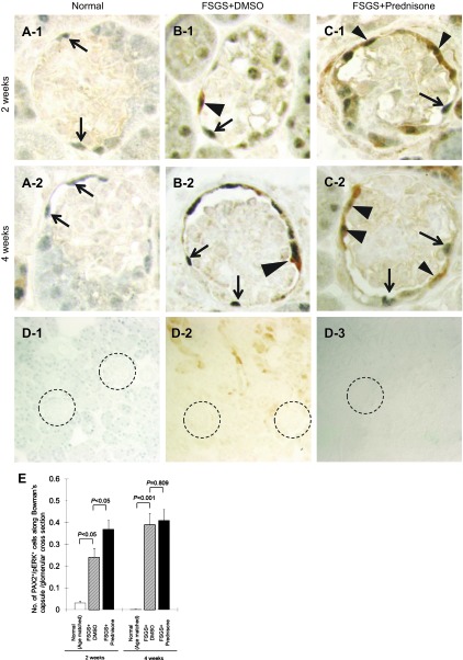
Fig. 8.
Prednisone augments p-ERK1/2 in parietal epithelial cells (PECs) in experimental FSGS. A–C: representative images of PAX2/p-ERK double staining at ×630 original magnification. The arrowheads show double positive cells along Bowman's capsule, arrows show PAX2 single positive cells. D-1 to D-3: staining was not detected when the primary antibodies were omitted as negative controls (×200). PAX2 only (D-1), p-ERK only (D-2), no primary antibody (D-3). Glomeruli are circled for easier identification. E: number of PECs with phosphorylated ERK, measured as the number of PAX2/p-ERK double positive cells/glomerulus along Bowman's capsule, increased at 2 and 4 wk in mice with FSGS mice (hatched bar). Prednisone treatment was associated with an increase in this number at 2 wk (filled bar).

