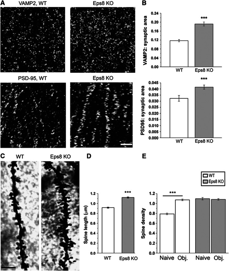
Figure 2.
Excessive synaptic growth and spine abnormalities in the hippocampus of Eps8 KO mice. (A, B) Representative fields of the CA1 hippocampal region of a wt and Eps8 KO mouse brains, stained for the synaptic vesicle protein synaptobrevin/VAMP2 and the glutammatergic postsynaptic protein, PSD-95. Quantitation of either pre- or postsynaptic areas reveals a larger number of synaptic contacts in Eps8 KO hippocampus. Scale bar, 5 μm. (C) Details of CA1 apical dendrites from wt and Eps8 KO hippocampi stained with Golgi-Cox technique. Scale bars, 10 μm. (D, E) Quantitation of spine length and density in wt or KO animals under control conditions (naïve) or after application of the novel object recognition test (obj) (length, D, wt spines=0.91±0.010 μm; KO spines=1.12±0.012 μm; total number of examined spines: 551, wt and 506, Eps8 KO; number of independent experiments: 3) (density, E, wt naive=0.789±0.017 spines per μm of parent dendrite; wt obj.=1.07±0.016 spines per μm; KO naive=1.09±0.024 spines per μm; KO obj.=1.08±0.016 spines per μm total number of examined dendritic branches: 321 wt ctr, 242 wt obj, 105 KO ctr, 361 KO obj; number of independent experiments: 3). Note that Eps8 KO mice display denser and longer spines, which fail in undergoing further increase in number after the object recognition test. Mann–Whitney rank sum test P<0.001. All data are expressed as mean±s.e.m. Six animals for each condition have been analysed.

