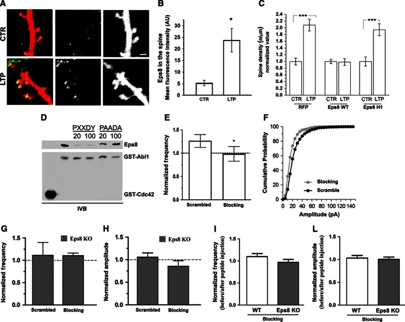
Figure 5.
The acute inhibition of Eps8 actin-capping activity precludes potentiation. (A) Representative examples of dendrites of mice hippocampal neurons transfected with RFP, exposed to chemical LTP and stained for Eps8. Note that Eps8 immunoreactivity at the spine head increases after potentiation. Scale bar, 2 μm. (B) Quantitation of Eps8 immunofluorescence at the spine head in vehicle-treated and glycine-treated (100 μM) neurons (Mann–Whitney Rank Sum Test, P=0.013) (total number of examined neurons: 16 untreated neurons and 12 gly-treated neurons; number of independent experiments: 3). (C) Quantitation of spine density under the different experimental conditions shows that potentiation is prevented by overexpression of Eps8 but not by its actin-capping mutant (total number of examined neurons: 15 ctr and 18 LTP for RFP, 29 ctr and 25 LTP for Eps8 wt and 18 ctr and 24 LTP for Eps8 H1; number of independent experiments: 5). (D) The proline rich consensus site of Abi1 (PXXDY) competes with Abi1 for binding to Eps8. Equal amounts of His-Eps8 (0.2 μM) were incubated with 0.2 μM immobilized GST-Abi1 in the absence or in the presence of 20 and 100 μM of either PXXDY or PAADA synthesized peptides. 2 μM GST-Cdc42 was used as a control. Proteins were analysed by immunoblotting with the indicated antibodies. (E, F) mEPSC frequency (E) and amplitude (F) in neurons exposed to chemical LTP and intracellularly perfused via the patch pipette with either the scrambled or the blocking peptide (the blocking peptide competes with Abi1 for binding to Eps8 and therefore inhibits the Eps8 capping activity). Note that neurons intracellularly perfused with the blocking peptide (grey column, white dots) are defective in potentiation, measured as mEPSC frequency or amplitude. Synaptic potentiation occurs in neurons intracellularly perfused with a scramble peptide (black dots) (total number of examined neurons: 6 for both conditions; number of independent experiments: 3). (G, H) mEPSC frequency (G) and amplitude (H) in Eps8 KO neurons exposed to chemical LTP and intracellularly perfused with either the scrambled or the blocking peptide. Note that mEPSC frequency and amplitude of KO neurons do not change upon glycine administration with or without injection of the blocking peptide. (I–L) Normalized mEPSC frequency (I) and amplitude (L) in WT and KO neurons before and after blocking peptide injection. Note that injection of the blocking peptide does not affect per se basal synaptic activity.

