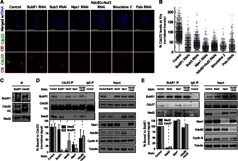
Figure 6.
Mps1-dependent BubR1 hyperphosphorylation is required for recruitment of Cdc20 to kinetochores and MCC formation. (A) Immunolocalization of Cdc20. Selected kinetochore pairs in the boxed areas are shown at higher-magnification. (B) Distribution of Cdc20 levels at unattached kinetochores for the experiments in (A). Cdc20 fluorescence intensities at kinetochores were determined relative to CID. n>20 cells for each condition. (C) Immunoprecipitation of Cdc20 and BubR1 from control cells lysates were probed by immunoblottimg for the indicated proteins. (D, E) Immunoprecipitation of Cdc20 (D) and BubR1 (E) from total cell lysates obtained from cells depleted of the indicated proteins. Immunoprecipitates (IP) and corresponding total cell lysates (Input) were probed by immunoblotting for the indicated proteins. Quantification of relative levels of protein bound to Cdc20 (C) or BubR1 (D) is shown. Co-immunoprecipitated protein signals were adjusted to the corresponding input signal and normalized for immunoprecipitated Cdc20 or BubR1. Graph bars in Cdc20 IP represent mean±s.d. and were obtained from three independent experiments. Values obtained for Cdc20 and BubR1 IP in control cells were set to 100%. Graph bars in BubR1 IP result from a single IP experiment. (A–E) Cultured cells were treated with MG132 for 1 h followed by 2 h of colchicine incubation. Binucleine 2 was added to cultures 30 min before microtubule depolymerization. Scale bars represent 5 μm.
Source data for this figure is available on the online supplementary information page.

