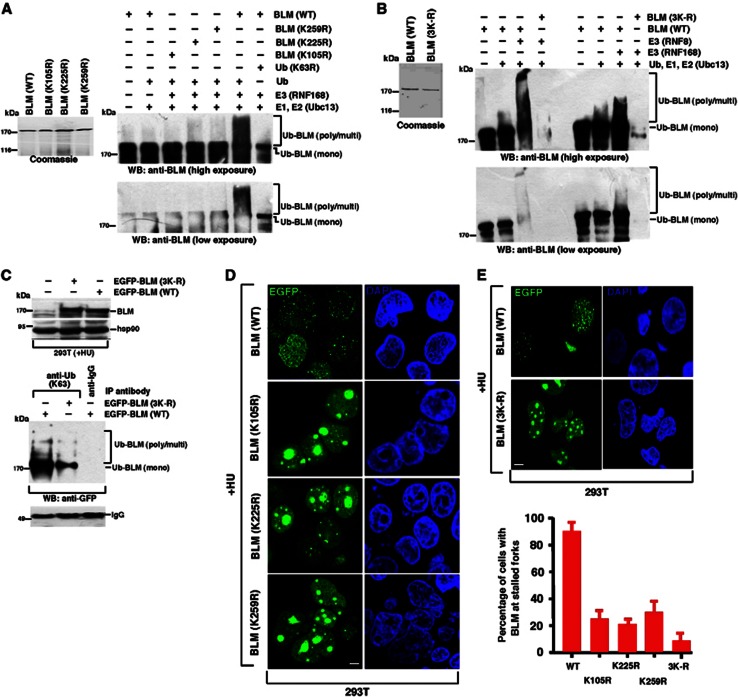
Figure 4.
Ubiquitylation of BLM at 105, 225 and 259 is required for BLM recruitment to the sites of stalled replication. (A) BLM undergoes K63-linked ubiquitylation at lysines residues 105, 225 and 259. (Left) Coomassie gel demonstrating the expression of GST-tagged BLM (WT) or BLM (K105R), BLM (K225R), BLM (K259R). (Right) In vitro ubiquitylation reactions were carried out using equal amounts wild-type BLM or the three BLM mutants (K105R, K225R and K259R). Western blots were carried out with antibodies against BLM (A300-120A). Two different exposures are shown to demonstrate the differential ubiquitylation. (B) Complete abrogation of RNF8-/RNF168-mediated ubiquitylation in BLM (3K-R) mutant. Same as (A) except BLM (3K-R) mutant was used. Expression of wild-type GST-tagged BLM and BLM (3K-R) mutant via Coomassie staining is shown on the left. RNF8 or RNF168 was used as the E3 ligase in parallel reactions. Two different exposures are shown to demonstrate the differential ubiquitylation. (C) Mutation of lysines at 105, 225 and 259 on BLM leads to loss of BLM poly-ubiquitylation after DNA damage. Wild-type BLM or (3K-R) mutant was overexpressed in 293T cells and subsequently treated with HU. (Top) The expression levels were determined by western analysis using antibodies against BLM (A300-110A) and hsp90. (Bottom) Immunoprecipitations were carried out using anti-K63-linked ubiquitin antibody. Immunoprecipitates were probed with antibodies against GFP or the corresponding IgG. Equal amount of antibody used for immunoprecipitation is demonstrated by comparing the IgG level. (D) Ubiquitylation of BLM at lysines 105, 225 and 259 is required for its recruitment to the stalled replication forks. 293T cells were transfected with EGFP-tagged wild-type BLM or BLM (K105R), BLM (K225R), BLM (K259R). Post-transfection the cells were treated with HU for 24 h. Transfected cells were tracked. Nuclei are stained by DAPI. Scale 5 μM. (E) Mutation of lysines at 105, 225 and 259 on BLM leads to enhanced nucleolar accumulation of BLM after HU treatment. (Top) Same as (D) except after transfection with either BLM (WT) or BLM (3K-R), the transfected cells were tracked. Nuclei are stained by DAPI. Scale 5 μM. (Bottom) Quantitation of (D) and (E, top).
Source data for this figure is available on the online supplementary information page.

