
Fig. 1.
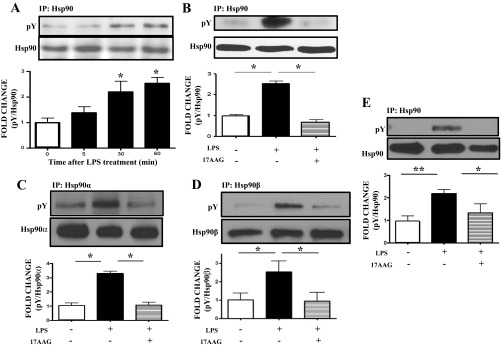
Modulation of pY levels of heat shock protein 90 (Hsp90) by LPS and 17-AAG. A: Western blot analysis of the expression levels of tyrosine-phosphorylated Hsp90 after 5-, 30-, or 60-min treatment with LPS in bovine pulmonary arterial endothelial cells (BPAEC). B: Western blot analysis of tyrosine-phosphorylated Hsp90 levels in BPAEC treated with LPS or vehicle and pretreated with PP2 or vehicle. C: Western blot analysis of tyrosine-phosphorylated Hsp90α levels in human lung microvascular endothelial cells (HLMVEC) treated with LPS or vehicle and pretreated with 17-AAG or vehicle. D: Western blot analysis of tyrosine-phosphorylated Hsp90β levels in HLMVEC treated with LPS or vehicle and pretreated with 17-AAG or vehicle. E: Western blot analysis of tyrosine-phosphorylated Hsp90 levels in lungs from mice treated with LPS or vehicle and pretreated with 17-AAG or vehicle. In all panels, Western blotting was performed on samples immunoprecipitated with antibodies against Hsp90 (A, B, E), Hsp90α (C), or Hsp90β (D), and each blot shown is representative of 3 separate experiments per group. Band intensity was analyzed by densitometry and normalized to Hsp90. *P < 0.05 vs. control, **P < 0.01 vs. control. Bars represent SE.