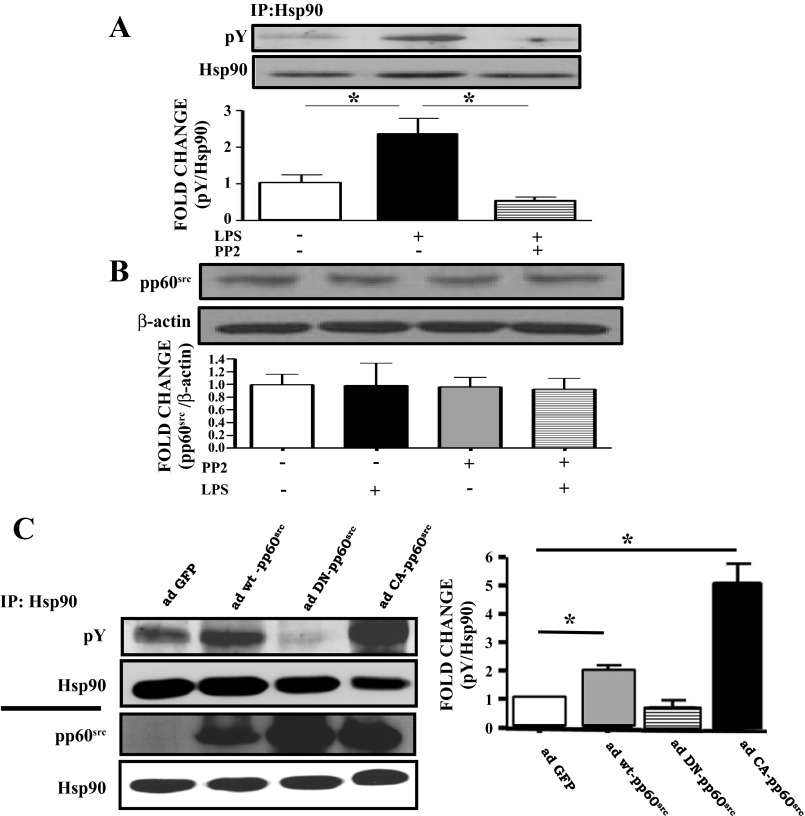
Fig. 3.
The pp60src inhibitor PP2 blocks LPS-induced Y phosphorylation of Hsp90, whereas constitutively active ad-pp60src increases Hsp90 Y phosphorylation in BPAEC. A: Western blot analysis of pY Hsp90 levels in BPAEC treated with LPS or vehicle and pretreated with PP2 or vehicle. Band density of pY was analyzed by densitometry and normalized to Hsp90 expression. *P < 0.05 vs. control. Western blotting was performed on samples immunoprecipitated with antibodies against Hsp90. B: Western blot analysis of pp60src expression in BPAEC treated with LPS or vehicle and pretreated with PP2 or vehicle. Band density of pp60src was analyzed by densitometry and normalized to β-actin. C: Western blot analysis of the expression of pY in BPAEC transduced with adenoviruses containing GFP (ad-GFP), wild-type pp60src (ad-wt-pp60src), dominant-negative pp60src (ad-DN-pp60src), or constitutively active pp60src (ad-CA-pp60src). Band density of pY was analyzed by densitometry and normalized to Hsp90. *P < 0.05 vs. ad-GFP. Western blotting was performed on samples immunoprecipitated with antibodies against Hsp90 (top). Western blot analysis of the expression of pp60src and Hsp90 in BPAEC transduced with ad-GFP, ad-wt-pp60src, ad-DN-pp60src and ad-CA-pp60src. Band density of pp60src was analyzed by densitometry and normalized to Hsp90. *P < 0.05 vs. ad-GFP (right). In all cases, each blot shown represents 1 of 3 independent experiments. Bars represent SE.

