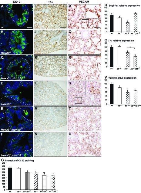
Fig. 3.
Characterization of lung epithelium and microvasculature of E18.5 Hoxa5;Hoxb5 mutant embryos. A–F: as assessed by immunofluorescence (IF), CC10 expression was decreased in lung airways of embryos carrying 2 Hoxa5 mutant alleles indicating the predominant role of Hoxa5 in club cell specification. G: mean CC10 intensity within the airway epithelium was measured by use of ImageJ. Values represent the average fluorescence intensity of the bronchial epithelial areas (surrounded by a dotted line). At least 4 pictures per specimen from 3–5 specimens were measured for each genotype. Values are expressed as means ± SE. I–N: immunostaining with podoplanin (T1α), a marker of type I pneumocytes, was considerably decreased in lungs from Hoxa5−/−, Hoxa5−/−;Hoxb5+/− and Hoxa5−/−;Hoxb5−/− embryos. P–U: PECAM-1 immunostaining revealed an undeveloped vascular network embedded in the thick lung mesenchyme in Hoxa5−/−, Hoxa5−/−;Hoxb5+/−, and Hoxa5−/−;Hoxb5−/− embryos. Scale bars: 100 μm (I–N), 50 μm (A–F, P–U). H, O, V: qRT-PCR analysis for Scgb1a1, T1α, and Vegfa expression in lungs from WT, Hoxa5−/−, Hoxb5−/−, and Hoxa5−/−;Hoxb5−/− E18.5 embryos. Comparison was made against WT. O: T1α expression was also significantly reduced in Hoxa5−/−;Hoxb5−/− specimens compared with Hoxa5−/− samples. Values are expressed as means ± SE. *P < 0.05, **P < 0.01, ***P < 0.001.

