Fig. 2.
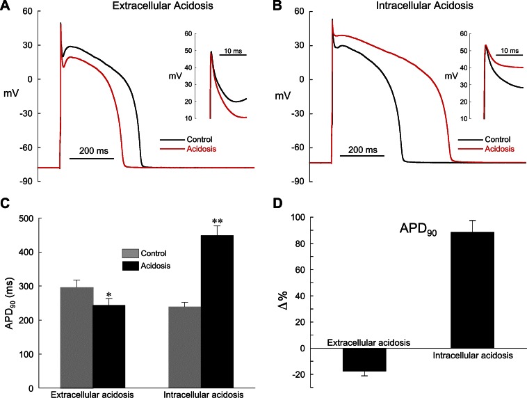
Effect of extra- and intracellular acidosis on epicardial action potentials (APs). A: extracellular acidosis (pHo 6.5) shortened AP duration (APD). Inset: expanded view of phase 1 repolarization. B: intracellular acidosis (80.0 mM acetate) prolonged APD, markedly slowed phase 1 repolarization, and nearly eliminated the notch. Inset: expanded view of phase 1 repolarization. C: summarized changes in APD at 90% repolarization (APD90) in low pHo (n = 5) and low pHi (n = 11). D: changes (Δ) in APD90 shown in C expressed as % relative to control. The normal pipette filling solution was used in these experiments (no BAPTA). Pacing cycle length (CL) for all experiments was 2 s. *P < 0.05, **P < 0.01, paired, control vs. acidosis.

