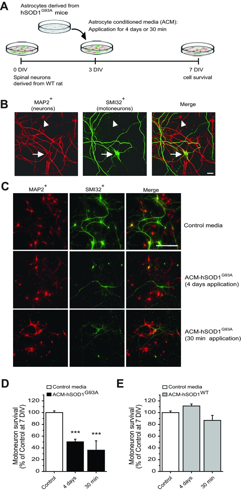
Fig. 1.
Long-term and short-term treatment of primary spinal cultures with ACM-hSOD1G93A equally trigger cell death of motoneurons. A: flow diagram of experiment. Medium was conditioned for 7 days by astrocytes derived from transgenic mice overexpressing human (h)SOD1G93A (ACM-hSOD1G93A). Primary wild-type (WT) rat spinal cord cultures (3 DIV) were exposed to ACM-hSOD1G93A for 4 days or for 30 min; all were fixed at 7 DIV to assay cell survival with immunocytochemistry. B: fixed 7 DIV primary spinal cultures were double-labeled with anti-microtubule-associated protein 2 (MAP2) antibody (red) to visualize interneurons (arrowhead) and motoneurons (arrow) and with the SMI-32 antibody (green) to identify motoneurons (arrow). Scale bar, 25 μm. C: spinal cultures were treated with control medium for 4 days (top) or ACM-hSOD1G93A for either 4 days (middle) or 30 min (bottom), fixed at 7 DIV, and labeled with MAP2 and SMI-32. Note that a single, short-term 30-min exposure of spinal cord neurons to ACM-hSOD1G93A is as effective in triggering motoneuron cell death as chronic application. Interneurons are spared in both conditions. Scale bar, 200 μm. D: graph of the ratio of SMI-32+/MAP2+ neurons showing % of surviving motoneurons at 7 DIV after treatment with control medium or ACM-hSOD1G93A applied for 4 days or 30 min. E: % of surviving motoneurons at 7 DIV after 4-day or 30-min application of 3 DIV cultures of medium that was conditioned by astrocytes derived from transgenic mice overexpressing hSOD1WT (ACM-hSOD1WT). Values represent means ± SE from at least 3 independent experiments performed in duplicate, analyzed by 1-way ANOVA followed by a Tukey post hoc test. ***P < 0.001 relative to control medium at 7 DIV.

