Fig. 5.
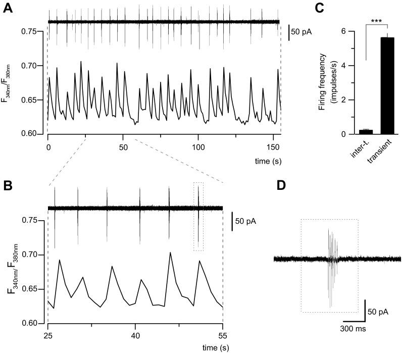
Calcium transients in cultured spinal cord neurons correlate with AP firing. A: simultaneous recording of spontaneous AP firing in cell-attached mode (top) and calcium transients (bottom) in a spinal cord neuron. B: expanded timescale for the neuron in A. Note the coincidence of calcium transients with bursts of AP firing. C: bar graph showing the mean frequency of AP firing from 198 transients recorded in 4 cultured spinal cord neurons. Note that during intertransient (inter-t.) periods, AP firing is virtually absent. Values represent means ± SE. ***P < 0.001, t-test. D: action currents in an expanded timescale from the last burst in B (dotted box).

