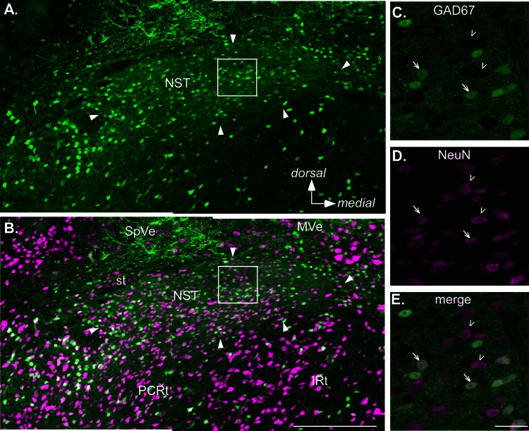
Fig. 1.
Confocal images of a section from a level of the rostral nucleus of the solitary tract (rNST) in an adult GAD67+ mouse approximately midway between the rostral pole of the nucleus and where the NST abuts the IVth ventricle. Sections were also immunostained for the neuronal marker NeuN. A and B: low-power maximum-intensity projections showing the distribution of GAD67+, enhanced green fluorescent protein (EGFP)-stained neurons and fibers (green, A) and EGFP expression merged with NeuN staining (magenta, B). Arrowheads indicate the borders of the nucleus. Note, however, that the most medial pole contains sparse somal label for either marker and corresponds with a region occupied by preganglionic parasympathetic neurons that constitute the rostral pole of the dorsal motor nucleus of the vagus. Aside from this region, GAD67+ neurons are distributed throughout the nucleus; likewise, the nucleus is characterized by profuse EGFP staining of the neuropil. C–E. higher-magnification images at a single z-plane. Arrows indicate examples of double-stained neurons; arrowheads indicate neurons stained only with NeuN. Scale bars, 250 μm (A and B), 25 μm (C–E). IRt, intermediate subdivision of the medullary reticular formation; MVe, medial vestibular nucleus; PCRt, parvocellular reticular formation; SpVe, spinal vestibular nucleus; st, solitary tract.

