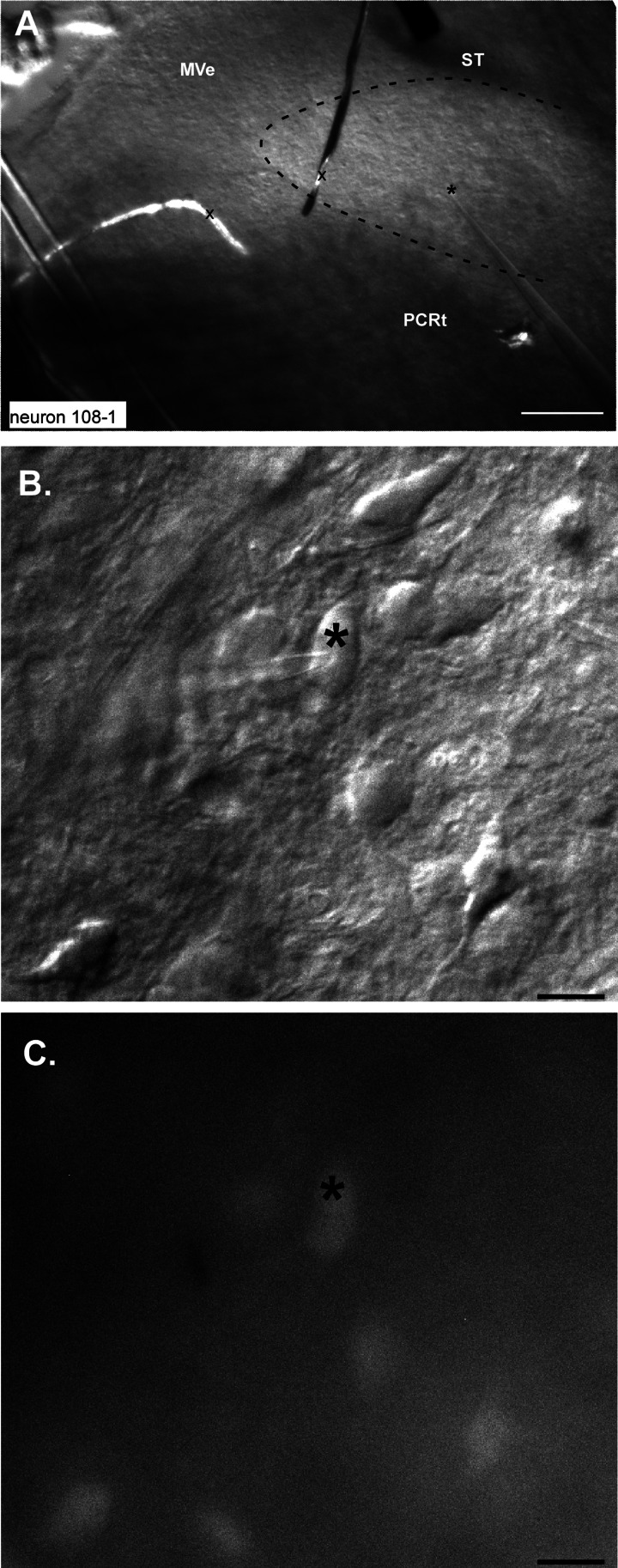
Fig. 3.
Photomicrographs of a ventrally located site where a GAD67+ neuron was recorded. A: low-magnification DIC image showing the stimulating electrode on the solitary tract (ST) and the recording electrode in the ventral 1/3 of the nucleus; the position of the tip is indicated by *. The approximate outlines of the rNST are indicated by the dashed line. Pieces of debris (x) cut across the medial part of rNST and medial vestibular nucleus (MeV). PCRt, parvocellular reticular formation. B: high-magnification DIC image showing the pipette on the recorded neuron. C: high-magnification fluorescent image showing that the cell is GAD67+. Scale bars, 250 μm (A), 25 μm (B and C).

