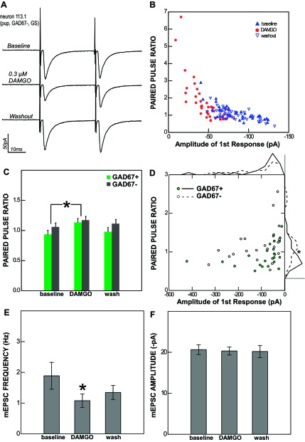
Fig. 5.
The effect of DAMGO on rNST neurons is presynaptic. A: the response of an rNST neuron to ST stimulation (averages of 40 sweeps in voltage clamp) before, during, and after the application of 0.3 μM DAMGO. B: single data points showing the relationship between the amplitude of the 1st response to ST stimulation and the paired-pulse ratio (PPR) for the same cell in each condition (baseline, DAMGO, and washout). C: across the population, the average PPR increases during DAMGO application for both GAD67+ and GAD67− neurons. *P < 0.001. D: scatterplot of average baseline amplitude and PPR values for individual monosynaptic ST-evoked responses (n = 44) showing an inverse relationship between the 1st response amplitude and PPR for both GAD67+ and GAD67− neurons. The PPRs of monosynaptic responses to ST stimulation were significantly smaller in GAD67+ neurons than in GAD67− neurons. This effect may be seen most clearly in the frequency polygons (binned in 15 equal segments) displayed outside the graph area; scale bars correspond to a frequency of 10 neurons. E: the frequency of miniature excitatory postsynaptic current (mEPSCs; tested in 7 neurons, 2 GAD67+ and 5 GAD67−) was significantly reduced by DAMGO. *P < 0.014. F: the average amplitude of mEPSCs was unaffected by DAMGO. Taken together, these results indicate a presynaptic site of action.

