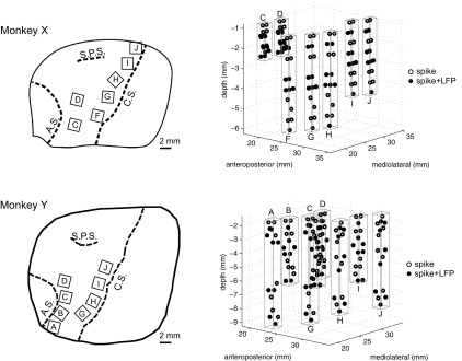
Fig. 2.
Neural recording locations. Simultaneous spiking and local field potential (LFP) activity was recorded from multiple floating microarrays (FMAs) in primary motor cortex (M1) and dorsal (PMd) and ventral (PMv) premotor areas of 2 subjects (monkeys X and Y). Left: drawings traced from intraoperative photos show the location of implanted arrays in each monkey, relative to cortical sulci (C.S., central sulcus; A.S., arcuate sulcus; S.P.S. superior precentral sulcus). Only the arrays used in the present study are shown. Right: neural recording sites from electrodes of staggered (i.e., nonuniform) length are indicated by circles. Open circles identify spike recording sites, and filled circles identify simultaneous spike and LFP recording sites. Spikes were acquired from every electrode, while LFPs were acquired from every other electrode. Rectangular prisms outlined in gray outline indicate the approximate recording volume sampled by each FMA.

