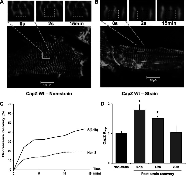
Fig. 1.
Time course of CapZβ1 dynamics after cyclic mechanical strain. Neonatal rat ventricular myocytes (NRVMs) infected by CapZβ1-GFP were either unstrained or subjected to 1-Hz cyclic 10% strain for 1 h. A and B: confocal images of CapZβ1-GFP with FRAP shown before bleach (0 s), after bleach (2 s), and 15 min later in cells in the following conditions: A, unstrained with resting spontaneous beating or B, 1 h after cyclic strain has ended. Region of interest (ROI) shown as dashed white boxes (3.75 μm × 3.75 μm). Inset: higher magnification images of area delineated by the solid white boxes in the lower magnification image. C: fluorescence recovery after photobleaching (FRAP) of CapZβ1-GFP as a percentage of prebleach intensity with unstrained control (dashed line, Non-S) and within 1 h after cyclic strain ended [solid line, S (0–1 h)]. D: Kfrap values for CapZβ1 normalized to unstrained cells; 0 to ∼1 h (n = 5), 1 to ∼2 h (n = 4), and 2 to ∼3 h (n = 4). Values are means ± SE. *Significant difference (P < 0.01).

